
Microscopes
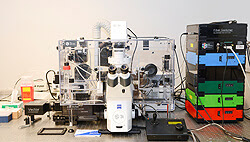
3i Spinning Disk Confocal using Slidebook software:
This is an inverted microscope platform capable of imaging with speeds up to 2,000 frames/sec. for high speed 3D cell imaging of live or fixed cells. Solid state lasers are coupled through the pinholes of the spinning disk to provide strong excitation light and high signal to noise images. FRAP and TIRF can be done on this system. The software synchronizes with the machine to provide maximum signal with minimum photobleaching.
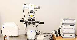
Zeiss LSM 700 Confocal using Zen software:
This laser scanning 3D microscope captures defined optical height sections of a sample and combines them in a 3D image stack. An image can be provided that shows topography quality and good edge detection. The software facilitates techniques such as FRAP,FRET, or Flip. The systems sensitivity permits fast, specimen preserving scanning in multiple fluorescence work.
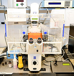
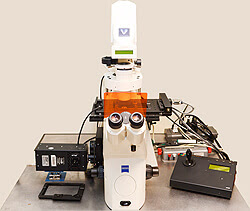
Live Cell Marianas:
(10th floor Goodman Building)
This inverted microscope is intended for live cell applications. It allows for rapid filter changes as well as changes that are controlled by filter wheels. It is fitted with a live cell chamber to control temperature, carbon dioxide levels and humidity. It has added filters for doing calcium flux experiments.
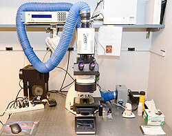
L W D Marianas:
(10th floor Goodman Building)
This inverted microscope uses dry objectives. It is especially suitable for cell culture observation plus phase detection.
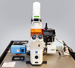
3i Marianas:
(5th floor Smith Building)
This microscope is equipped with a filter wheel to control excitation and emission light and a motorized x-y stage.
Leica DMRXA using Slidebook software:
This is an upright wide field microscope using 4 color digital deconvolution and DIC fluorescence. The system is optimized for quantitative, high resolution, high-resolution, three dimensional analysis of multi-labels in fixed cells and tissue sections. It uses a filter wheel at the light source.