
Quantitative Imaging Laboratory
About Us
The Quantitative Imaging Laboratory (QIL) at National Jewish Health (NJH), directed by Stephen Humphries PhD and David Lynch MB, focuses on development and application of the latest medical image analysis methods in clinical and translational research. The QIL was established in 2008 to support radiologic imaging in the COPDGene research study and provide image archival and analysis for the NJH Department of Radiology. Today, the QIL’s main goal is to develop and apply novel quantitative image analysis methods, including those based on Artificial Intelligence (AI), that have meaningful clinical impact.
 The QIL also provides quality assurance and image data management and is currently the primary image analysis core for local, national and international multicenter imaging research studies including: COPDGene, which includes 10,000 patients and nearly 20,000 CT exams, and the Pulmonary Fibrosis Foundation Registry which aims to create an imaging directory consisting of CT images of over 1,000 subjects.
Research Activities
The QIL develops novel techniques to recognize and characterize normal and abnormal anatomy, researches methods for efficient large-scale processing of medical images and provides quantitative image analysis for research and clinical purposes. The QIL is actively engaged in research on deep learning, a class of AI algorithms that use multi-layer computer models called neural networks to “learn” from example data rather than being explicitly programmed.
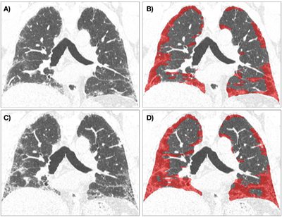

Researchers in the QIL have developed deep learning programs that are capable of automatic detection and quantification of pulmonary fibrosis on CT scans. The Data-Driven Texture Analysis (DTA) algorithm developed by the QIL correlates with radiologist visual assessment and pulmonary function, is sensitive to change in extent of fibrosis, and can predict survival. Quantitative analysis of lung fibrosis has been applied in multiple completed and ongoing clinical trials of treatment in fibrotic lung disease.
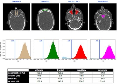
The QIL has developed deep learning methods for automatic 3D segmentation of the nasal sinuses on CT scans. This enables quantitative assessment of sinus volumes and the extent of sinus opacification as a marker of sinusitis. Recent research indicates a correlation between CT sinusitis scores and asthma diagnosis, eosinophil count and pulmonary function. Support from the State of Colorado has facilitated QIL research and participation in a multicenter trial for severe nasal polyposis.
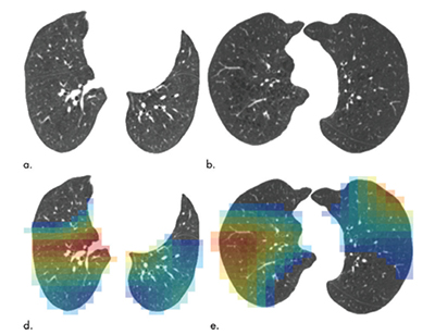
The QIL is the imaging core for the COPDGene study; imaging in COPDGene has resulted in over 100 publications and several important advances, summarized in a recent review article by Bhatt et al. The QIL has been particularly interested in the role of CT in classifying emphysema, and in the potential of deep learning to assist with this task. Emphysema pattern has been shown to be an important indicator of disease severity but is time consuming and subjective to perform visually. The QIL has developed a deep learning algorithm to automatically score emphysema severity pattern on CT according to a classification system developed by the Fleischner Society. Further research includes quantitative phenotyping of emphysema, air trapping, and airway analysis.
Faculty
 Stephen M. Humphries, PhD, Director and Imaging Scientist
Associate Professor in the Department of Radiology at National Jewish Health, is a medical physicist and imaging scientist with a major interest in quantitative CT of fibrotic and obstructive diffuse lung diseases.
As the Director of the Quantitative Imaging Laboratory at National Jewish Health, Dr. Humphries oversees all laboratory efforts including quality assurance, storage, and image analysis for multi-center CT studies of COPD, pulmonary fibrosis and rhinosinusitis.
 David Lynch, MD, Medical Director
Professor of Radiology at National Jewish Health, principal radiology investigator for the imaging core of COPDGene (which has enrolled over 10,000 individuals with and without COPD, now followed for over 10 years), and IPFNet. He has authored or coauthored over 300 papers and several books on lung imaging, He is Past President of the Society of Thoracic Radiology and of the Fleischner Society.